Lambda Virus Microscope / Bacteriophage Lambda Early Pioneer And Still Relevant Sciencedirect / Fever, stress, sunlight can trigger viral genes to take over cells 3.
Lambda Virus Microscope / Bacteriophage Lambda Early Pioneer And Still Relevant Sciencedirect / Fever, stress, sunlight can trigger viral genes to take over cells 3.. ▲ the new coronavirus under the electron microscope. Latent virus infections • usually, virus is limited by defense system • herpes virus infection (cold sores/fever blisters) 1. Basic optical microscopes can be very simple, although many complex. The virus was isolated from a patient in the u.s. Elements of the lambda life cycle.
Here's what we know about how dangerous it is, and how well vaccines work against it. Over 5500 bacterial viruses have so far been characterized by electron microscopy, making bacteriophages, at least on paper, the largest viral group in existence. The corona virus is rna group of virus which contains rna as its genetic material. The lambda genome is 48.5 kb (compared to that of t4 which is 172 kb). The infected animal tissue can be prepared for examination with an electron microscope 17.
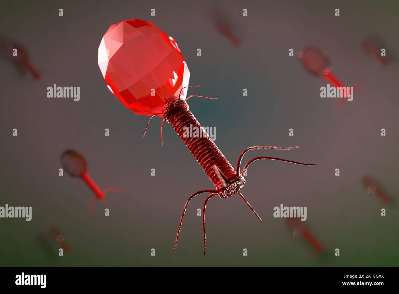
Bacteriophage High Resolution Stock Photography And Images Alamy from c8.alamy.com The infected animal tissue can be prepared for examination with an electron microscope 17. Optical microscopes are the oldest design of microscope and were possibly invented in their present compound form in the 17th century. Over 5500 bacterial viruses have so far been characterized by electron microscopy, making bacteriophages, at least on paper, the largest viral group in existence. Elements of the lambda life cycle. Basic optical microscopes can be very simple, although many complex. Lambda phage or coliphage λ is a bacteriophage that infects the bacteria belonging to the members of the bacterial species escherichia coli ().; (ii) which virus contains dna? And is seen here emerging from.
A bacteriophage (/ b æ k ˈ t ɪər i oʊ f eɪ dʒ /), also known informally as a phage (/ ˈ f eɪ dʒ /), is a virus that infects and replicates within bacteria and archaea.the term was derived from bacteria and the greek φαγεῖν (phagein), meaning to devour.bacteriophages are composed of proteins that encapsulate a dna or rna genome, and may have structures that are either.
The lambda variant was reportedly discovered in peru. The lambda variant might be more transmissible and be strong enough to avoid vaccines. Even with the highest numerical aperture (na) objective lens (theoretical max of 1.00 f. Resembles t4producing factory, and the cell soon lyses and but only has a singlereleases its viral products. Infected person always harbors the virus in cells 2. The infected animal tissue can be prepared for examination with an electron microscope 17. The principal contribution of electron microscopy to bacteriophage research is the technique of negative staining. Basic optical microscopes can be very simple, although many complex. Here's what we know about how dangerous it is, and how well vaccines work against it. These mutations may make it easier for lambda to bind to our cells and make it harder for our antibodies to latch onto the virus and neutralize it. If you are trying to look at small order scales of dna and chromatin, then there is no chance. The lambda phage was originally discovered by esther lederberg in 1951 in the us during her studies on e. Bacteriophage models or types 1.
If you are trying to look at small order scales of dna and chromatin, then there is no chance. A) ebola b) hiv c) λ (lambda) phage d) tobacco mosaic (iii) explain why an electron microscope, rather than a light microscope, was used to produce this photograph. (ii) which virus contains dna? Lambda phage or coliphage λ is a bacteriophage that infects the bacteria belonging to the members of the bacterial species escherichia coli ().; Even with the highest numerical aperture (na) objective lens (theoretical max of 1.00 f.
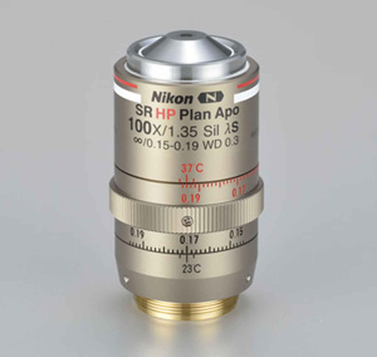
New Objective Lens For Biological Microscope Cfi Sr Hp Plan Apochromat Lambda S 100xc Sil News Nikon Europe B V from d33b8x22mym97j.cloudfront.net The infected animal tissue can be prepared for examination with an electron microscope 17. A bacteriophage (/ b æ k ˈ t ɪər i oʊ f eɪ dʒ /), also known informally as a phage (/ ˈ f eɪ dʒ /), is a virus that infects and replicates within bacteria and archaea.the term was derived from bacteria and the greek φαγεῖν (phagein), meaning to devour.bacteriophages are composed of proteins that encapsulate a dna or rna genome, and may have structures that are either. The optical microscope, also referred to as a light microscope, is a type of microscope that commonly uses visible light and a system of lenses to generate magnified images of small objects. If you are trying to look at small order scales of dna and chromatin, then there is no chance. ▲ the new coronavirus under the electron microscope. Lambda phage, lamb and peg will be flowed into the space between two cover slips under a fluorescence microscope to trigger and observe the partial ejection of lambda genome. And is seen here emerging from. Lambda phage or coliphage λ is a bacteriophage that infects the bacteria belonging to the members of the bacterial species escherichia coli ().;
The optical microscope, also referred to as a light microscope, is a type of microscope that commonly uses visible light and a system of lenses to generate magnified images of small objects.
And is seen here emerging from. To test our hypothesis that transcription of n drives genome delivery, the length of genome ejected will be measured before and after transcription with and without protein n. Even with the highest numerical aperture (na) objective lens (theoretical max of 1.00 f. Lambda phage, lamb and peg will be flowed into the space between two cover slips under a fluorescence microscope to trigger and observe the partial ejection of lambda genome. These mutations may make it easier for lambda to bind to our cells and make it harder for our antibodies to latch onto the virus and neutralize it. Lambda phage or coliphage λ is a bacteriophage that infects the bacteria belonging to the members of the bacterial species escherichia coli ().; The corona virus is rna group of virus which contains rna as its genetic material. The lambda phage was originally discovered by esther lederberg in 1951 in the us during her studies on e. Here's what we know about how dangerous it is, and how well vaccines work against it. The lambda genome is 48.5 kb (compared to that of t4 which is 172 kb). Infected person always harbors the virus in cells 2. Latent virus infections • usually, virus is limited by defense system • herpes virus infection (cold sores/fever blisters) 1. Basic optical microscopes can be very simple, although many complex.
These mutations may make it easier for lambda to bind to our cells and make it harder for our antibodies to latch onto the virus and neutralize it. Elements of the lambda life cycle. The corona virus is rna group of virus which contains rna as its genetic material. Light microscopes are severely limited in their resolving power. An electron microscope is similar to an optical microscope.
Decision Making At A Subcellular Level Determines The Outcome Of Bacteriophage Infection Cell from els-jbs-prod-cdn.jbs.elsevierhealth.com Infected person always harbors the virus in cells 2. If you are trying to look at small order scales of dna and chromatin, then there is no chance. Elements of the lambda life cycle. The principal contribution of electron microscopy to bacteriophage research is the technique of negative staining. Here's what we know about how dangerous it is, and how well vaccines work against it. Resembles t4producing factory, and the cell soon lyses and but only has a singlereleases its viral products. Fever, stress, sunlight can trigger viral genes to take over cells 3. Cells are destroyed, cold sore develops burton's microbiology:
The optical microscope, also referred to as a light microscope, is a type of microscope that commonly uses visible light and a system of lenses to generate magnified images of small objects.
The lambda variant was reportedly discovered in peru. Fever, stress, sunlight can trigger viral genes to take over cells 3. The lambda phage was originally discovered by esther lederberg in 1951 in the us during her studies on e. Cells are destroyed, cold sore develops burton's microbiology: The optical microscope, also referred to as a light microscope, is a type of microscope that commonly uses visible light and a system of lenses to generate magnified images of small objects. The principal contribution of electron microscopy to bacteriophage research is the technique of negative staining. The corona virus is rna group of virus which contains rna as its genetic material. A bacteriophage (/ b æ k ˈ t ɪər i oʊ f eɪ dʒ /), also known informally as a phage (/ ˈ f eɪ dʒ /), is a virus that infects and replicates within bacteria and archaea.the term was derived from bacteria and the greek φαγεῖν (phagein), meaning to devour.bacteriophages are composed of proteins that encapsulate a dna or rna genome, and may have structures that are either. Resembles t4producing factory, and the cell soon lyses and but only has a singlereleases its viral products. Lambda phage or coliphage λ is a bacteriophage that infects the bacteria belonging to the members of the bacterial species escherichia coli ().; Latent virus infections • usually, virus is limited by defense system • herpes virus infection (cold sores/fever blisters) 1. These mutations may make it easier for lambda to bind to our cells and make it harder for our antibodies to latch onto the virus and neutralize it. Elements of the lambda life cycle.


Posting Komentar
0 Komentar